Stanford researchers have developed a microscope system that generates high-quality images in low-light conditions, and preserves delicate samples. Light can damage sensitive microscopy samples, which often leads researchers to use low-light conditions that produce low-quality images. Unlike conventional microscope systems, this multi-pass microscopy system passes a low-light beam through a sample multiple times. The sample’s image reflects back onto itself multiple times and generates increasing contrast and detail with each cycle. The resulting high contrast image has improved resolution of phase or absorption differences, and is compatible with low-light conditions - preserving the sample without sacrificing data quality. The approach can be applied to beams of light or electrons, in transmission or reflection, and in dark-field or bright-field configuration.
Stage of ResearchLaboratory proof-of-concept is complete. High resolution prototype design is underway.
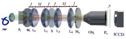
Multi-pass Microscopy System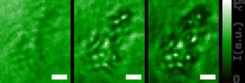
Multi-pass microscopy images of embryonic kidney 293T cells – Repeated phaseshifts and absorption lead to an increased visibility of the cells. The scale bar is 20 μm.