Stanford researchers have developed a simple and non-toxic method for more streamlined and precise electron beam radiotherapy using 3D printed electron field shaping devices. These 3D-printed cutouts can provide more accurate electron radiotherapy with reduced toxicity, labor, and cost compared to traditional Cerrobend methods.
3D printed designs for electron cutout have been demonstrated to accurately reproduce the dose profiles compared to that of Cerrobend cutouts. This method removes toxic material from the clinic, reduces manual labor, and provides improved reproduction of the field placement and field shape compared to Cerrobend. Given the current rapid rate of development of 3D printing, it is expected that these technologies will be dramatically improved in the coming years, giving yet more convenience, speed, and precision. This increased precision, in concert with other recent developments, such as modulating electron bolus, opens up new opportunities for advancing electron radiotherapy.
Figure 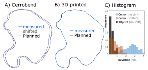
Figure description - Fig 1. 3D printed cutouts provide more accurate field shaping.
(A) The planned (black line) and the measured Cerrobend outlines (blue line) have a mean deviation of 2.6±0.2 mm. Even after shifting, the Cerrobend outline (dashed line) shows a maximum deviation of 2 mm and a mean of 0.8 mm compared to the planned outline. (B) The 3D printed cutout (blue line) follows the planned outline (black line) more closely with maximum deviation of 1 mm and a mean of 0.4 mm. (C) Histogram of deviations between final and planned field shapes.
Stage of Research- Prototype tested
- Demonstrated that 3D-printed cutouts can provide more accurate electron radiotherapy with reduced toxicity compared to traditional Cerrobend methods
Related Technologies:Stanford docket S19-098
"Quality Assurance (QA) Phantom for off-axis spatial accuracy for frameless single-isocenter Multitarget Stereotactic Radiosurgery"